Intraoperative skeletal traction in the dog
G. L. Rovesti, A. Margini Veterinary Clinic M. E. Miller, Cavriago, Reggio Emilia, Italy
F. Capp ellari, B. Peirone Department of Animal Pathology, School of Veterinary Medicine of Turin, Grugliasco, Turin, Italy
Introduction
In veterinary medicine, intra-operative skeletal traction has not been commonly used for fracture reduction (1, 3, 5, 6), despite its widespread application and significant benefits in human orthopaedic surgery. In human trauma patients, standardized, reproducible techniques are routinely employed for fracture reduction. These techniques include proper patient positioning, specific instrumentation, and application of skeletal traction (4, 8). A recent cadaveric study established the critical parameters for skeletal traction in dogs that were analogous to those utilized in humans (7). In the present study, we evaluated the clinical efficacy of skeletal traction for intra-operative fracture reduction in dogs with appendicular diaphyseal fractures.
Material and methods
The animals included in this study were client-owned dogs with traumatic fractures that were treated by the authors in the period from January 2000 to January 2003 at the Veterinary Clinic ‘M. E. Miller’ in Cavriago or the School of Veterinary Medicine of Turin Small Animal Clinic. For each animal, data were collected for: signalment, source of trauma, clinical history, circumference of the anatomical region treated, type of fracture, interval between the occurrence of the fracture and the surgical intervention, the modality of traction, and the method of fracture stabilization.
The circumference of the fractured region was determined with a flexible measuring tape. Measurements were made at the level of the axilla for the humerus, the middiaphysis for the antebrachium, the inguinal region for the femur, and the distal portion of the tibial crest for the tibia. In cases in which the fracture was located at one of these predetermined points, measurements were taken immediately distal or proximal to the fracture.
Application of skeletal traction
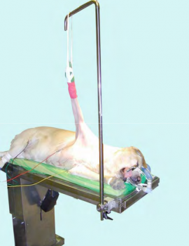
Skeletal traction was used for the reduction of fractures using equipment (Ergomed 99, Med Matrix, Modena, Italy), opposition points and anchorage points as previously described by us (7). The anchorage points used for application of traction was a traction stirrup attached to a transcondylar Kirschner wire in the relevant humerus or femur (Figs. 1, 2) and the anchorage belts for the antebrachium and tibia (Fig. 3). During the application of traction, the maximal traction load was measured using a dynamometer (Yo- Zuri America, Lucie, FL, USA). With the goal of avoiding iatrogenic tissue injury, the maximum load applied to each limb was never allowed to exceed 25 kg. The duration of traction was also recorded. and was included as part of the surgical time, even when traction was initiated before limb disinfection and preparation of the operating theatre.
The traction modalities varied in each case, based on whether the fracture was closed or open, and depending on its location. Usually, the dogs that had closed fractures of the antebrachium and tibia were positioned on the traction table; traction being applied before the limb was scrubbed. Once the fracture segments were deemed to be realigned, fracture reduction was confirmed by digital palpation, fluoroscopy, or a combination of both methods. In this setting, the entire procedure, including the application of increasing traction loads needed to achieve the fracture reduction, was performed by the surgeon in charge. Once the fracture was satisfactorily realigned, the limb was maintained in traction, scrubbed and prepared for surgery in the normal manner. The traction devices were considered to be non-sterile, and were not included in the surgical field. For closed fractures of the humerus and femur, the limb was first scrubbed and prepared for surgery. After making the surgical approach to the fracture, a sterile traction stirrup was applied to the condyle (7), and then connected to the micrometric traction stand with a small sterile chain. The end of this chain connected to the stirrup was kept sterile, while the end connected to the dynamometer and distraction stand became contaminated. The load required to distract the fracture segments was applied by a non-scrubbed operating room assistant, who increased the load at the surgeon’s request.This was done by turning the handle at the top of the traction device in a counter clockwise direction, hence increasing the length of the device. When a decrease in tension was necessitated – the handle was turned in a clockwise direction. Contamination of the surgical field was avoided because the assistant could manipulate the traction device at its end, far from the surgical field, while the portion of the device close to the surgical field remained covered with sterile towels (Fig. 1).
Correction of angular malalignment of fractures was performed entirely by the nonscrubbed assistant who manoeuvred the traction device under the direction of the surgeon, as described previously (7). For the correction of procurvatum and recurvatum malalignment, the assistant unlocked the clamp securing the traction device, allowing the surgeon to slide it along the lateral rail on the table. Once the malalignment had been corrected, the clamp was re-locked in the new position.All of the open fractures were managed in sterile conditions from the beginning of the procedure, irrespective of their location.
Evaluation of fracture reduction and alignment
After fracture reduction, all of the fractures were stabilized by the application of external skeletal fixation, bone plate or locked nailing. Radiographs, taken immediately after the surgical procedure, were used to evaluate the contact between the fracture fragments and the axial realignment of the treated limb using the following scale:
- Excellent: 90% to 100% contact between fracture fragments and axial malalignment in any plane of less than 5°;
- Good: 50% to 89% contact between fracture fragments and axial malalignment in any plane of less than 10°;

Fig. 1 Operating theatre showing traction application for treatment of a femoral fracture (case 4).
- Fair: 10% to 49% contact between fracture fragments and axial malalignment in any plane of less than 30°;
- Poor: 0 to 9% contact between fracture fragments and axial malalignment in any plane of 30° or more.
In comminuted fractures, the only feature of the fracture reduction taken into consideration by the surgeon was axial alignment, since it was not possible to realise fragment contact.
Data analysis
A Pearson’s correlation test was performed to evaluate correlation between limb diameter and load applied, and between latency time and the load applied. Dogs were arbitrarily divided into two groups, based on whether latency time to surgery was less than three days, or three days and longer.
The differences between these two groups in the maximum traction load, and in the duration of traction were evaluated using the Student t-test. Values of P < 0.05 were considered significant.
Results
The animals included in this study were 21 dogs of various breeds (13 males, eight females) (Table 1). Three animals had ultiple fractures.

Fig. 2 Same case as in Fig. 1, showing anchorage of the traction stirrup to the supracondylar region of the distal femur (case 4).
The average age was three years (range 0.5 to 9) and the average body weight was 23.7 kg (range 5 to 45). All of the fractures were diaphyseal, without any articular component, and were classified as being transverse (n=10), oblique (n=4) or comminuted (n=10). Four of the fractures were open (Table 2), and were grade I or II in severity (2). Overall, the average maximum traction load applied was 18kg weight (range 8 to 25), while the average duration of traction was 48 min (range 10 to 150). Data for limb circumference, latencytime, traction load, traction time, and surgery time for each fracture location are summarized in Table 2.A correlation was not detected between limb diameter and load applied (r = – 0.03), and there was a poor correlation between latency time and load applied (r = 0.3). The differences between the two groups in maximum traction load (kg weight) (17 ± 5 versus 18 ± 5) was not significant (P = 0.613). However, the difference between the two groups in traction duration (minutes) (29 ± 16 versus 58 ± 32) was significant (P = 0.02).

Fig. 3 Preoperative phase, showing traction application by means of anchorage belts for reduction of radiusulna fracture (case 6).

Once the desired reduction had been achieved, the application of osteosynthesis implants was greatly simplified in that the osseous segments were maintained in correct alignment for the necessary amount of time. In particular, internal fixation could be carried out utilizing a reduced number of bone clamps, which permitted easier application of the osteosynthesis plate (Fig. 4). In many of the comminuted fractures, the bone fragments that were widely displaced from the bone axis, spontaneously repositioned themselves following traction, probably due to compressive centripetal forces exerted by muscles subjected to traction. The insertion of external skeletal fixator pins and subsequent frame construction were also simplified (5) (Fig. 5). Neither anchorage belts, nor traction stirrup hindered the surgical procedure. For traction of the antebrachium and tibia, the anchorage belts were positioneddistal to the fracture, around the metacarpus or metatarsus. For traction of the humerus and femur, the stirrup was positioned on the condyle, thus allowing full surgical approach to the bone itself (Figs. 1, 2, 4). In two of the humeral fractures, fracture reduction by traction was only achieved after the application of a second Kirschner wire and traction stirrup to the proximal end of the humerus (Fig. 6). This approach was adopted because the initial attempt at humeral traction with a single distal stirrup caused significant distal translation of the scapula, without obtaining satisfactory alignment of the humeral fracture.
In fracture treatment of the distal segments,it was possible to first obtain fracture reduction, and then prepare the limb for surgery. This approach allowed the surgeon more freedom with the reduction manoeuvres, especially with leverage, than would have been possible during conventional osteosynthesis procedures. Furthermore, the technique also allowed less experienced surgeons to perform the fracture stabilization, once a more experienced surgeon had obtained its correct reduction and realignment.
Osteosynthesis was carried out in 11 cases using plates and screws, in ten cases with external skeletal fixation, and in three cases with locked nails. Postoperative alignment of the 24 treated segments was judged as excellent in 21 fractures and good in the remaining 3 fractures. Two complications were observed. In one case, a gap of 3 mm between the fracture ends of a humerus treated with a locked nail was seen on the postoperative radiographs. The second complication occurred in a Newfoundland dog with a femoral fracture, in which the contralateral limb developed oedema distal to the stifle a few days after surgery. This was presumed to have been due to compression caused by the opposition belt surrounding the inguinal area, and it resolved without further complication within a few days.

Fig. 4 Reduction of a femoral fracture obtained with skeletal traction alone (case 1).

Discussion
In this study we found that the manoeuvres for skeletal traction aimed at reducing diaphyseal fracture were rather straightforward, and intra-operative axial deviations
were easily resolved by moving the traction bar as previously described (7). This system of application of skeletal traction for fracture reduction has some elasticity that is inherent to the animal’s tissue and the anchoring and opposition bands, that renders the process non-linear during the initial stages. Although the application of opposition and anchorage belts is relatively simple, slippage of these belts may also contribute to this problem (7) or result in local tissue injury. On the other hand, the traction applied
via a traction stirrup resulted in negligible elastic drop and did not cause any compressive soft tissue injury.

Fig. 5 Pin insertion for treatment of radius-ulna fracture by external skeletal fixation. Reduction is maintained by skeletal traction (case 8).
With just one exception, we believe that it is important to use the opposition points that were developed from our cadaver study (7), and to monitor the duration and magnitude of the loading force in order to avoid any tissue damage. The findings of the present clinical study suggest that opposition bands may not be sufficient to counteract skeletal traction applied to the humerus because they do not prevent the entire scapula from being pulled distally. Althoughthe traction forces (17–25 kg weight) used to achieve reduction of humeral fractures were similar to those employed in the other bones, we found that it was necessary to place a traction stirrup on the proximal end of the humerus in two of the fractures. Subsequent to completion of the present study, this has become our standard technique for application of skeletal traction in humeral fractures in the clinical setting.
Excessive traction could also potentially result in a compromising of the nervous and vascular systems. In our case series, such a complication occurred in one dog who developed a limb oedema. In circumstances in wich an elevated load must be applied, it might be prudent to minimize its duration in order to reduce the likelihood of complications. When the procedure can not be completed in a sufficiently brief period, it clamp or Kirschner wire in order to allow stabilization of the fracture with either a
clamp or Kirschner wire in order to allow the tissues to be better perfused, and then resume traction after a short period. Once the reduction and stabilization of the fracture are considered to be adequate, then the traction load may be reduced according to the surgeon’s judgment.

Fig. 6 Intra-operative phase, showing humeral traction with two opposing traction stirrups in order to avoid scapula translocation (case 4).
From the present series of cases, we found that the interval between trauma and surgical intervention seemed to have a significant effect on duration of traction needed for reduction, but not the maximum traction load. The latter is perhaps not too
surprising, since the maximum load applied to any limb was never allowed to exceed 25
kg weight. Furthermore, limb circumference, which could be considered to be a crude index of resistance of the muscular mass to fracture reduction, was not correlated with either the amount or duration of traction. The ease of reduction may be influenced by other factors, such as fracture comminution. Usually, in comminuted fractures angular alignment is the main feature considered for reduction evaluation, and a small degree of axial shortening of a long bone is not considered to be a clinical problem. A further evaluation of technique would be required to better define the factors involved. Specific criteria for case selection were not established. As a general rule, any fracture may be more easily reduced if subjected to traction, because traction counteracts uscle contraction and shortening. In our experience, very small dogs are more difficult to properly position than large dogs. Joint fractures may be subjected to traction, but the development of guidelines and techniques for the reduction reof such fractures were beyond the scope of the study presented herein.
In conclusion, the results of our study suggested that proper patient positioning and the use of skeletal traction are easily learned techniques that can rapidly become standard procedures. Although the time required for setting up the table, positioning of the patient, and performance of traction were somewhat lengthy, this time was regained during the osteosynthesis phase. In fact, the technique maintains the fracture ends in a stable position for the entire duration of the procedure. Due to the unavoidable fatigue of the surgical assistant, such conditions are not reproducible with conventional methods of manual fracture reduction. The technique also allowed for reduced manipulation of soft tissues, particularly those involving muscle bellies. In this way, surgical intervention can be carried out while respecting the ‘open but don’t touch’ philosophy, with potential advantages for healing (1). However, the technique may be potentially dangerous; therefore in order to avoid iatrogenic trauma it should be applied with caution. It is imperative that the application of opposition and anchorage points are correct, and that excessive and prolonged loading is avoided.


